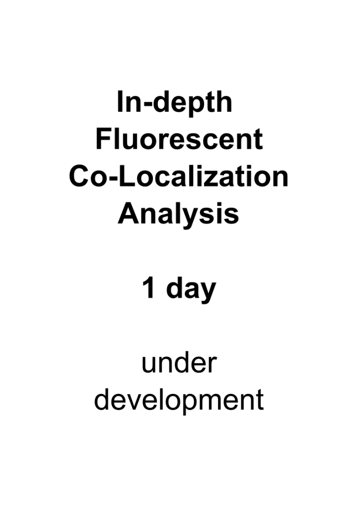
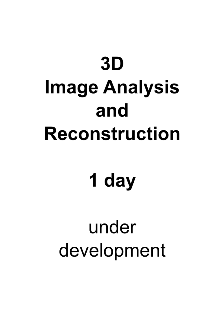
Workshop Overview
Click in the flyer images to get detailed information
Basic Level
Recommended start before the analysis course
Intermediate Level (required for advanced courses)
Advanced Level (the intermediate level analysis course is pre-requisite for those courses)
Workshop Calendar
June 2024
| Mon | Tue | Wed | Thu | Fri | Sat | Sun |
|---|---|---|---|---|---|---|
|
Saturday June 1
1
|
Sunday June 2
2
|
|||||
|
Monday June 3
3
|
Tuesday June 4
4
|
Wednesday June 5
5
|
Thursday June 6
6
|
Friday June 7
7
|
Saturday June 8
8
|
Sunday June 9
9
|
|
Monday June 10
10
|
Tuesday June 11
11
|
Wednesday June 12
12
|
Thursday June 13
13
|
Friday June 14
14
|
Saturday June 15
15
|
Sunday June 16
16
|
|
Monday June 17
17
|
Tuesday June 18
18
|
Wednesday June 19
19
|
Thursday June 20
20
|
Friday June 21
21
|
Saturday June 22
22
|
Sunday June 23
23
|
|
Monday June 24
24
|
Tuesday June 25
25
|
Wednesday June 26
26
|
Thursday June 27
27
|
Friday June 28
28
|
Saturday June 29
29
|
Sunday June 30
30
|
July 2024
| Mon | Tue | Wed | Thu | Fri | Sat | Sun |
|---|---|---|---|---|---|---|
|
Monday July 1
1
|
Tuesday July 2
2
|
Wednesday July 3
3
|
Thursday July 4
4
|
Friday July 5
5
|
Saturday July 6
6
|
Sunday July 7
7
|
|
Monday July 8
8
|
Tuesday July 9
9
|
Wednesday July 10
10
|
Thursday July 11
11
|
Friday July 12
12
|
Saturday July 13
13
|
Sunday July 14
14
|
|
Monday July 15
15
|
Tuesday July 16
16
|
Wednesday July 17
17
|
Thursday July 18
18
|
Friday July 19
19
|
Saturday July 20
20
|
Sunday July 21
21
|
|
Monday July 22
22
|
Tuesday July 23
23
|
Wednesday July 24
24
|
Thursday July 25
25
|
Friday July 26
26
|
Saturday July 27
27
|
Sunday July 28
28
|
|
Monday July 29
29
|
Tuesday July 30
30
|
Wednesday July 31
31
|
August 2024
| Mon | Tue | Wed | Thu | Fri | Sat | Sun |
|---|---|---|---|---|---|---|
|
Thursday August 1
1
|
Friday August 2
2
|
Saturday August 3
3
|
Sunday August 4
4
|
|||
|
Monday August 5
5
|
Tuesday August 6
6
|
Wednesday August 7
7
|
Thursday August 8
8
|
Friday August 9
9
|
Saturday August 10
10
|
Sunday August 11
11
|
|
Monday August 12
12
|
Tuesday August 13
13
|
Wednesday August 14
14
|
Thursday August 15
15
|
Friday August 16
16
|
Saturday August 17
17
|
Sunday August 18
18
|
|
Monday August 19
19
|
Tuesday August 20
20
|
Wednesday August 21
21
|
Thursday August 22
22
|
Friday August 23
23
|
Saturday August 24
24
|
Sunday August 25
25
|
|
Monday August 26
26
|
Tuesday August 27
27
|
Wednesday August 28
28
|
Thursday August 29
29
|
Friday August 30
30
|
Saturday August 31
31
|
September 2024
| Mon | Tue | Wed | Thu | Fri | Sat | Sun |
|---|---|---|---|---|---|---|
|
Sunday September 1
1
|
||||||
|
Monday September 2
2
|
Tuesday September 3
3
|
Wednesday September 4
4
|
Thursday September 5
5
|
Friday September 6
6
|
Saturday September 7
7
|
Sunday September 8
8
|
|
Monday September 9
9
|
Tuesday September 10
10
|
Wednesday September 11
11
|
Thursday September 12
12
|
Friday September 13
13
|
Saturday September 14
14
|
Sunday September 15
15
|
|
Monday September 16
16
|
Tuesday September 17
17
|
Wednesday September 18
18
|
Thursday September 19
19
|
Friday September 20
20
|
Saturday September 21
21
|
Sunday September 22
22
|
|
Monday September 23
23
|
Tuesday September 24
24
|
Wednesday September 25
25
|
Thursday September 26
26
|
Friday September 27
27
|
Saturday September 28
28
|
Sunday September 29
29
|
|
Monday September 30
30
|
October 2024
| Mon | Tue | Wed | Thu | Fri | Sat | Sun |
|---|---|---|---|---|---|---|
|
Tuesday October 1
1
|
Wednesday October 2
2
|
Thursday October 3
3
|
Friday October 4
4
|
Saturday October 5
5
|
Sunday October 6
6
|
|
|
Monday October 7
7
|
Tuesday October 8
8
|
Wednesday October 9
9
|
Thursday October 10
10
|
Friday October 11
11
|
Saturday October 12
12
|
Sunday October 13
13
|
|
Monday October 14
14
|
Tuesday October 15
15
|
Wednesday October 16
16
|
Thursday October 17
17
|
Friday October 18
18
|
Saturday October 19
19
|
Sunday October 20
20
|
|
Monday October 21
21
|
Tuesday October 22
22
|
Wednesday October 23
23
|
Thursday October 24
24
|
Friday October 25
25
|
Saturday October 26
26
|
Sunday October 27
27
|
|
Monday October 28
28
|
Tuesday October 29
29
|
Wednesday October 30
30
|
Thursday October 31
31
|
November 2024
| Mon | Tue | Wed | Thu | Fri | Sat | Sun |
|---|---|---|---|---|---|---|
|
Friday November 1
1
|
Saturday November 2
2
|
Sunday November 3
3
|
||||
|
Monday November 4
4
|
Tuesday November 5
5
|
Wednesday November 6
6
|
Thursday November 7
7
|
Friday November 8
8
|
Saturday November 9
9
|
Sunday November 10
10
|
|
Monday November 11
11
|
Tuesday November 12
12
|
Wednesday November 13
13
|
Thursday November 14
14
|
Friday November 15
15
|
Saturday November 16
16
|
Sunday November 17
17
|
|
Monday November 18
18
|
Tuesday November 19
19
|
Wednesday November 20
20
|
Thursday November 21
21
|
Friday November 22
22
|
Saturday November 23
23
|
Sunday November 24
24
|
|
Monday November 25
25
|
Tuesday November 26
26
|
Wednesday November 27
27
|
Thursday November 28
28
|
Friday November 29
29
|
Saturday November 30
30
|
December 2024
| Mon | Tue | Wed | Thu | Fri | Sat | Sun |
|---|---|---|---|---|---|---|
|
Sunday December 1
1
|
||||||
|
Monday December 2
2
|
Tuesday December 3
3
|
Wednesday December 4
4
|
Thursday December 5
5
|
Friday December 6
6
|
Saturday December 7
7
|
Sunday December 8
8
|
|
Monday December 9
9
|
Tuesday December 10
10
|
Wednesday December 11
11
|
Thursday December 12
12
|
Friday December 13
13
|
Saturday December 14
14
|
Sunday December 15
15
|
|
Monday December 16
16
|
Tuesday December 17
17
|
Wednesday December 18
18
|
Thursday December 19
19
|
Friday December 20
20
|
Saturday December 21
21
|
Sunday December 22
22
|
|
Monday December 23
23
|
Tuesday December 24
24
|
Wednesday December 25
25
|
Thursday December 26
26
|
Friday December 27
27
|
Saturday December 28
28
|
Sunday December 29
29
|
|
Monday December 30
30
|
Tuesday December 31
31
|
January 2025
| Mon | Tue | Wed | Thu | Fri | Sat | Sun |
|---|---|---|---|---|---|---|
|
Wednesday January 1
1
|
Thursday January 2
2
|
Friday January 3
3
|
Saturday January 4
4
|
Sunday January 5
5
|
||
|
Monday January 6
6
|
Tuesday January 7
7
|
Wednesday January 8
8
|
Thursday January 9
9
|
Friday January 10
10
|
Saturday January 11
11
|
Sunday January 12
12
|
|
Monday January 13
13
|
Tuesday January 14
14
|
Wednesday January 15
15
|
Thursday January 16
16
|
Friday January 17
17
|
Saturday January 18
18
|
Sunday January 19
19
|
|
Monday January 20
20
|
Tuesday January 21
21
|
Wednesday January 22
22
|
Thursday January 23
23
|
Friday January 24
24
|
Saturday January 25
25
|
Sunday January 26
26
|
|
Monday January 27
27
|
Tuesday January 28
28
|
Wednesday January 29
29
|
Thursday January 30
30
|
Friday January 31
31
|
February 2025
| Mon | Tue | Wed | Thu | Fri | Sat | Sun |
|---|---|---|---|---|---|---|
|
Saturday February 1
1
|
Sunday February 2
2
|
|||||
|
Monday February 3
3
|
Tuesday February 4
4
|
Wednesday February 5
5
|
Thursday February 6
6
|
Friday February 7
7
|
Saturday February 8
8
|
Sunday February 9
9
|
|
Monday February 10
10
|
Tuesday February 11
11
|
Wednesday February 12
12
|
Thursday February 13
13
|
Friday February 14
14
|
Saturday February 15
15
|
Sunday February 16
16
|
|
Monday February 17
17
|
Tuesday February 18
18
|
Wednesday February 19
19
|
Thursday February 20
20
|
Friday February 21
21
|
Saturday February 22
22
|
Sunday February 23
23
|
|
Monday February 24
24
|
Tuesday February 25
25
|
Wednesday February 26
26
|
Thursday February 27
27
|
Friday February 28
28
|
March 2025
| Mon | Tue | Wed | Thu | Fri | Sat | Sun |
|---|---|---|---|---|---|---|
|
Saturday March 1
1
|
Sunday March 2
2
|
|||||
|
Monday March 3
3
|
Tuesday March 4
4
|
Wednesday March 5
5
|
Thursday March 6
6
|
Friday March 7
7
|
Saturday March 8
8
|
Sunday March 9
9
|
|
Monday March 10
10
|
Tuesday March 11
11
|
Wednesday March 12
12
|
Thursday March 13
13
|
Friday March 14
14
|
Saturday March 15
15
|
Sunday March 16
16
|
|
Monday March 17
17
|
Tuesday March 18
18
|
Wednesday March 19
19
|
Thursday March 20
20
|
Friday March 21
21
|
Saturday March 22
22
|
Sunday March 23
23
|
|
Monday March 24
24
|
Tuesday March 25
25
|
Wednesday March 26
26
|
Thursday March 27
27
|
Friday March 28
28
|
Saturday March 29
29
|
Sunday March 30
30
|
|
Monday March 31
31
|
April 2025
| Mon | Tue | Wed | Thu | Fri | Sat | Sun |
|---|---|---|---|---|---|---|
|
Tuesday April 1
1
|
Wednesday April 2
2
|
Thursday April 3
3
|
Friday April 4
4
|
Saturday April 5
5
|
Sunday April 6
6
|
|
|
Monday April 7
7
|
Tuesday April 8
8
|
Wednesday April 9
9
|
Thursday April 10
10
|
Friday April 11
11
|
Saturday April 12
12
|
Sunday April 13
13
|
|
Monday April 14
14
|
Tuesday April 15
15
|
Wednesday April 16
16
|
Thursday April 17
17
|
Friday April 18
18
|
Saturday April 19
19
|
Sunday April 20
20
|
|
Monday April 21
21
|
Tuesday April 22
22
|
Wednesday April 23
23
|
Thursday April 24
24
|
Friday April 25
25
|
Saturday April 26
26
|
Sunday April 27
27
|
|
Monday April 28
28
|
Tuesday April 29
29
|
Wednesday April 30
30
|
No events.
May 2025
| Mon | Tue | Wed | Thu | Fri | Sat | Sun |
|---|---|---|---|---|---|---|
|
Thursday May 1
1
|
Friday May 2
2
|
Saturday May 3
3
|
Sunday May 4
4
|
|||
|
Monday May 5
5
|
Tuesday May 6
6
|
Wednesday May 7
7
|
Thursday May 8
8
|
Friday May 9
9
|
Saturday May 10
10
|
Sunday May 11
11
|
|
Monday May 12
12
|
Tuesday May 13
13
|
Wednesday May 14
14
|
Thursday May 15
15
|
Friday May 16
16
|
Saturday May 17
17
|
Sunday May 18
18
|
|
Monday May 19
19
|
Tuesday May 20
20
|
Wednesday May 21
21
|
Thursday May 22
22
|
Friday May 23
23
|
Saturday May 24
24
|
Sunday May 25
25
|
|
Monday May 26
26
|
Tuesday May 27
27
|
Wednesday May 28
28
|
Thursday May 29
29
|
Friday May 30
30
|
Saturday May 31
31
|
No events.
If you want to organize a workshop for a graduate program, contact BioVoxxel for group prices.
INDIVIDUALS CAN BOOK HERE OR VISIT THE TICKET SHOP FOR MORE INFO
Why education in scientific image handling, processing and analysis is important!
The immense publication volume in life sciences is constantly increasing. New methods including diverse imaging techniques and the further technical innovation and advance e.g. in modern microscopy contribute to faster data acquisition in progressively less time.
Nevertheless, drawing final conclusions from acquired data is still the responsibility of the individual scientists and working groups. In the light of this increasing acquisition speed and amount of data in today’s scientific environment it is indispensable to be aware of background knowledge regarding data processing and comply with scientific standards to obtain non-altered and meaningful data sets as bases of our interpretations. Moreover, every scientist should adhere to the respective scientific ethics and good laboratory practice to sustain the significance and credibility of scientific data in the future.
Most young students in life sciences right at the beginning of their scientific careers often are not fully aware of the possibilities, limits and problems of digitized imaging data. Therefore, it is essential to teach especially young academics in an early stage of their career a comprehensive knowledge about image handling, basic processing and analysis. This should combine an efficient workflow with high scientific quality in the future.
Moreover, one essential part in scientific publication is to present data in form of figures. Therefore, a least biased choice of representative images from an experiment is already the first critical step (besides proper experimental design and image acquisition). Thereafter, images are often subject to editing. The reason/intention for the image editing should already be questioned. Is it applied to improve visibility of features, to direct the observers view to specific regions, to get rid of “ugly background” or dirt from the sample preparation or to underscore points for or against a hypothesis?
Mostly, image editing (as well as image analysis) is done based on good intentions but influenced by natural bias (e.g. our visual system [1]) and often a lack of sound knowledge in image processing techniques. Not seldom, this leads to alterations in the image data which very quickly might be considered as misrepresentation of data. The Office of Research Integrity (ORI) figured out that from all cases they opened in 2007-2008 for investigation against potential scientific misconduct 68% included image data manipulation [2].
Of consideration is the fact that images we take in science ARE precious data and need to be handled as such [3].
In the last years the awareness in this field increased and the number of journals explicitly stating strict image data related guidelines and enforcing compliance is (too) slowly increasing.
Additionally, several whitepapers with a common tenor of suggested guidelines for image processing of scientific images are available [4], [5], [6], [7], [8].
Besides those guidelines the education of young scientists in image processing, analysis, handling of scientific images and publication ethics is essential. BioVoxxel is dedicated to communicate skills in good scientific practice regarding digital images to the scientific community to help preventing unintentional image data alterations.
Finally, the way of visually presenting data is important. Therefore, BioVoxxel offers workshops in basic Scientific Illustration to teach how to efficiently communicate data in graphical abstracts or schematic figures.
Scientific Figure Making with Fiji and Inkscape (I2K, Oct. 2023)
Schmied, C., Nelson, M.S., Avilov, S. et al. Community-developed checklists for publishing images and image analyses. Nat Methods 21, 170–181 (2024). https://doi.org/10.1038/s41592-023-01987-9
Figure Making Best Practices (BINA, March 2024)
Basic image processing and tools (ImageJ Conference 2015, Madison)
References
[1] Seeing the Scientific Image
[2] Science journals crack down on image manipulation
[3] Digital Images Are Data: And Should Be Treated as Such
[4] Guidelines for Best Practices in Image Processing
[6] Digital Image Ethics – University of Arizona (by Douglas Cromey)
[7] What’s in a picture? The temptation of image manipulation
[8] Manipulation and Misconduct in the Handling of Image Data
[9] Seeing the Big Picture – Scientific Image Integrity under Inspection


